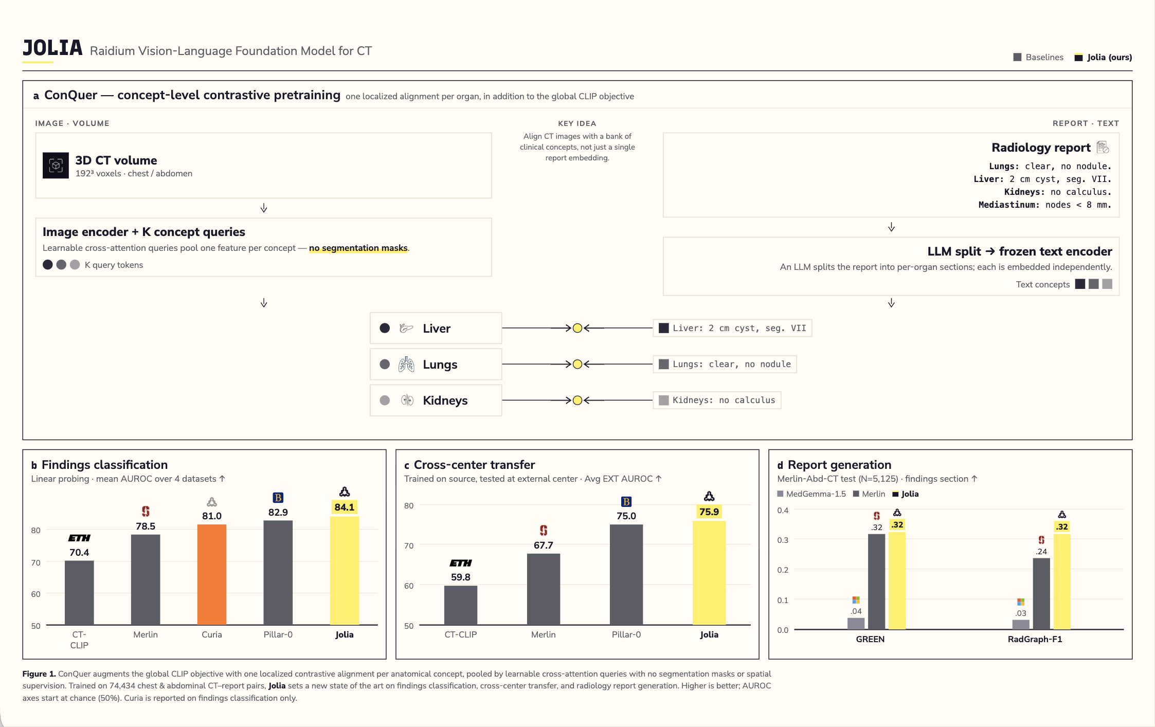
Jolia, our vision-language foundation model for chest and abdominal CT, set a new standard for ML research for radiology when we released it, pairing imaging with the clinical language radiologists use to describe it.
Today, we are introducing Jolia Pro the scaled successor to Jolia. Built on the same ConQuer method, it connects what the model sees in each part of a scan, to the precise words radiologists use to describe each organ in a report, rather than treating the report as a single block of text.
This new release leverages a larger backbone, increased compute, and an expanded training set combining public datasets with proprietary CT and report data. For a full walkthrough of the ConQuer method, read our original Jolia announcement
I. What is new
Jolia Pro grows the model's core from Jolia's 22M parameters to 80M, and scales training from a single node to 16. More importantly, it moves beyond public data alone: alongside the CT-RATE, INSPECT, and Stanford datasets used for Jolia, Jolia Pro adds two proprietary sources, one drawn from everyday hospital imaging and one from an additional oncology-focused abdominal collection.
II. What actually improved
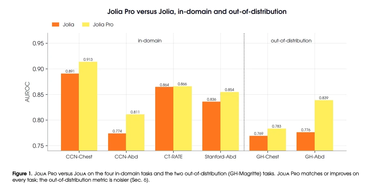
On the two public benchmarks that let us compare Jolia and Jolia Pro directly, on equal footing, Jolia Pro improves average AUROC (our standard score for how well the model tells abnormal scans from normal ones) by roughly one point. That gain is concentrated almost entirely in abdominal CT, with chest performance remaining essentially the same. That is largely by design: the public dataset Jolia was trained on already skewed heavily toward chest, and on top of that, more of the new proprietary data added was abdominal than chest, giving the model meaningfully more new abdominal signal to learn from overall.
Extracting that gain was not automatic: one proprietary source was itself chest-heavy, and mixing it without adjustment actually crowded out abdominal signal rather than adding it. Correcting for that with body-part-balanced sampling (making sure the model sees a fair mix of chest and abdomen scans during training, rather than whatever ratio the raw data happens to come in) mattered more than raw model size or compute. Abdomen is where we've added the most data so far, and where we expect see the most improvement as we keep expanding the dataset.
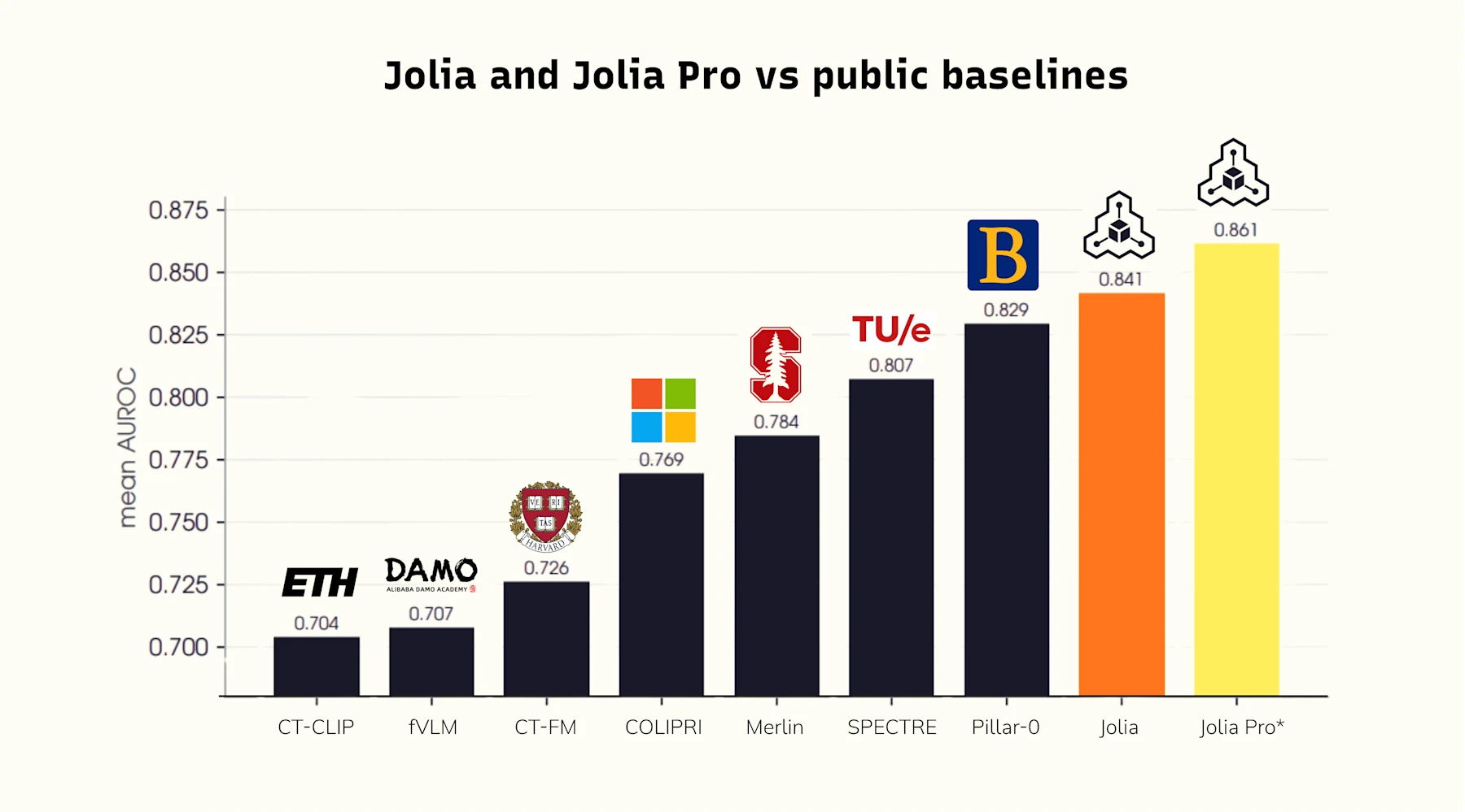
That same pattern holds when we test transfer to a center outside our training data: Jolia Pro's biggest improvement is in abdomen, with chest increasing slightly by 1.4 points, since it was already well covered. Jolia Pro also clearly outperforms the public baselines here, though we're treating these external numbers directionally for now, since only a narrow set of findings overlap between this benchmark and our own training taxonomy.
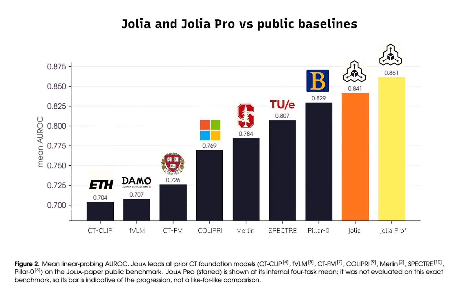
III. The more interesting finding: scale alone isn't the story
We ran a systematic test across model size, learning rate, batch size, node count, data mixture, and data augmentation to understand what actually drives improvement. Past a fairly modest model size, simply making the model bigger did not help on its own, and without adjusting the learning rate for that larger size, it made the results worse. What helped instead was more ordinary: tuning the learning rate to the model’s size, finding the right batch size, correcting data imbalance through balanced sampling, light data-quality filtering, and knowing when to stop training before it overfits.
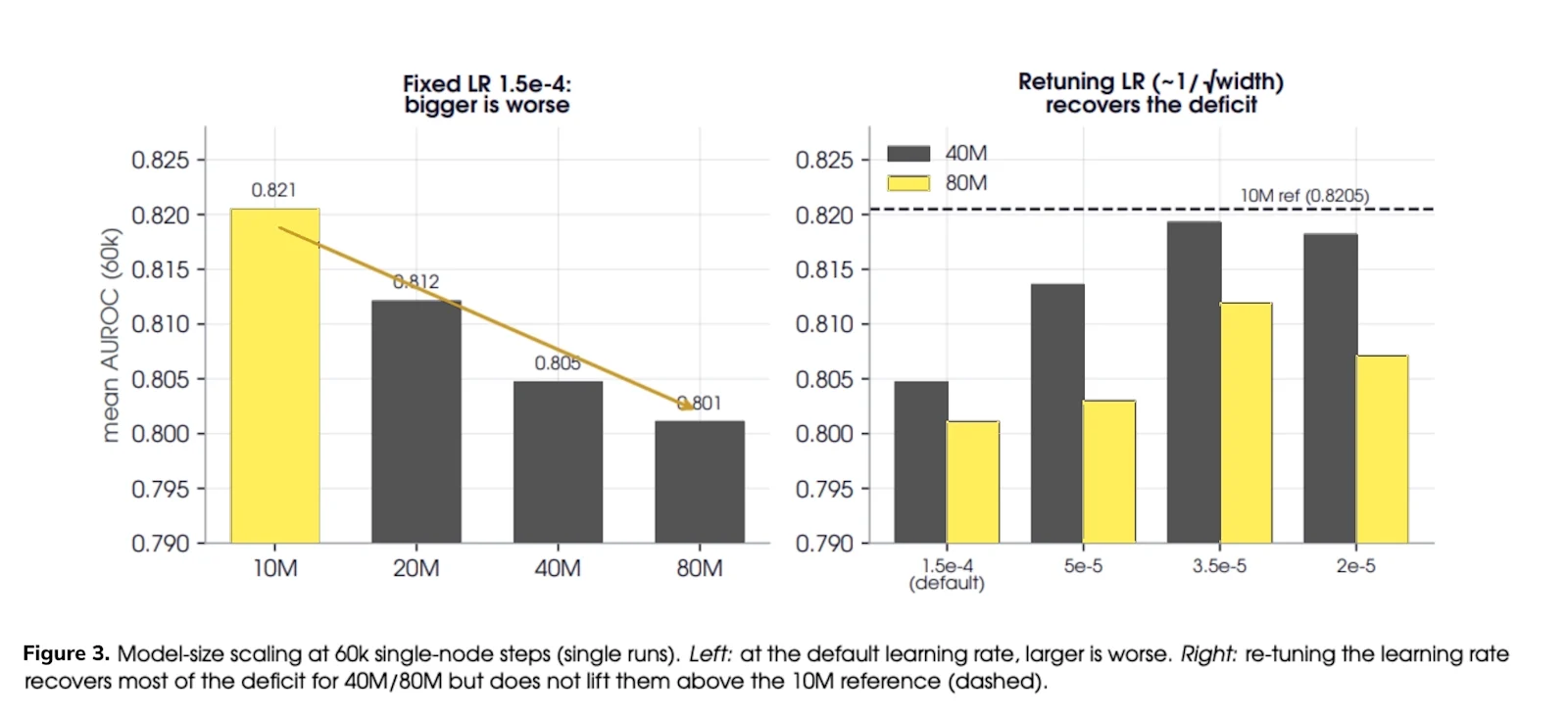
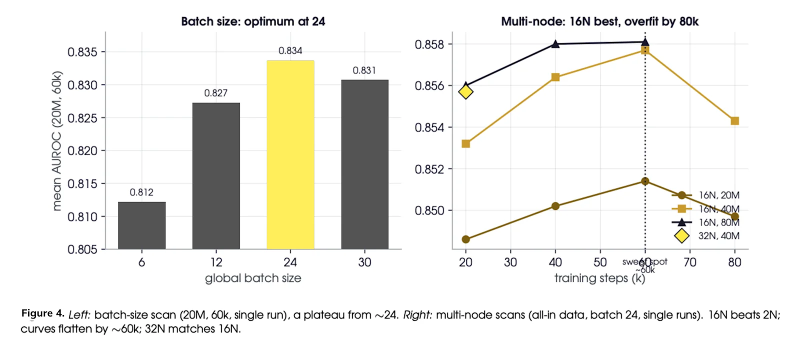
This is a useful signal for the field: continued improvement in 3D CT foundation models is not just about bigger models and more compute. It's about getting the training approach right at every step, which is exactly the kind of grounded, science-driven work we want to be known for.
IV. What this means going forward
Jolia Pro is the strongest model in the Jolia line to date. Where Curia and Curia-2 build structural understanding of a scan, the Jolia line puts that same discipline toward findings: connecting what the model sees to the language radiologists use to describe it. This will have a significant contribution to Raidium Read, the generalist viewer we are building, so this progress feeds directly into our product.
The clearest open problem ahead of us is closing the gap in out-of-distribution abdominal transfer (making sure the model performs just as reliably on scans from hospitals and scanners it wasn't trained on), and building a measurement protocol solid enough to report that gap with confidence. That's where the next iteration of this work is headed.
Jolia Pro's models and evaluation datasets are proprietary; get in touch if you'd like to learn more about how this fits into our roadmap.
Jolia and Jolia Pro are research models. Results shown are Raidium's own internal evaluations and are not independently peer reviewed.
Ready to explore Jolia?
Read our Jolia preprint on arXiv
Download our Jolia model on Huggingface
Contact us to access Jolia Pro model